Consultation and analysis
Skin analysis with SkinVision: making sense of images, scores and measurements
Skin analysis is most useful when the practitioner can explain what each result represents. SkinVision helps organise facial images and reports, while a clear distinction between an image score and a physical measurement keeps the consultation accurate and understandable.
What does SkinVision bring to a consultation?
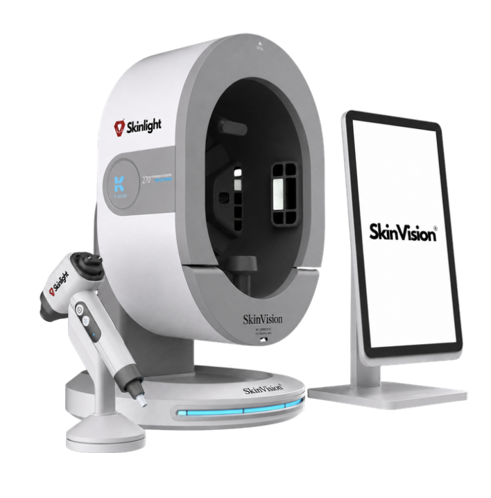
SkinVision captures facial images using several types of illumination and supports comparison and reporting. SkinVision DUO extends the consultation scope to facial and scalp imaging. The images can help a practitioner point out visible characteristics, document a baseline and review changes at later visits. [3] [4]
Different lighting modes emphasise different features. Software then analyses aspects of the captured image. An output may describe the apparent area, intensity or distribution of a feature, or place it within an algorithmic scoring system. Explaining the method is more helpful than presenting every number as an absolute biological fact. [1] [3]
How is an image score different from a measurement?
An image-derived score should be described as an assessment of the image. It is not a direct measurement of collagen quantity, the amount of melanin in physical units, or transepidermal water loss. A percentage or letter grade on a report does not change the underlying measurement method. [1] [2]
Hydration is a useful example. Dedicated probes can assess electrical properties associated with water in the outer skin. The Corneometer, for instance, uses capacitance and reports its own instrument units. Where a SkinVision configuration includes a separate hydration probe, its reading should be identified by the actual probe method and kept distinct from image scores. Confirm the supplied accessory and reporting scale. [2]
Keep comparisons consistent
Research on another facial imaging system, VISIA, has examined the reproducibility and accuracy of wrinkle and estimated skin-age outputs. This is useful background for asking how an imaging result was obtained and validated. It does not validate SkinVision's algorithms or make scores from different brands interchangeable. [1]
For your clinic, use a repeatable approach to photography. Keep positioning, lighting mode and the area being compared consistent. Record relevant changes such as skincare use or recent treatment. A visible difference between two photographs is easier to interpret when the conditions under which they were captured are understood.
Explain the result in plain language
A useful explanation starts with the observation: this lighting mode makes an area easier to see, and this score summarises the software's analysis of that image. Then discuss how the observation fits the client's concerns and the practitioner's assessment. Give the client an opportunity to ask what a label or colour scale means.
Avoid turning an estimated skin age into a diagnosis or a promise of how quickly a treatment will work. Images support a consultation; concerns that require medical assessment need the appropriate referral. A clear report can remain valuable without claiming that it measures everything about the skin.
- Label image scores and separate probe measurements clearly.
- Compare the same areas and capture modes at follow-up.
- Record observations, the agreed plan and questions that need further assessment.
Make the report useful after the visit
Agree which photographs and report details are relevant to the consultation and how the client can access them. Within the clinic, use consistent naming so that another authorised team member can understand the record without having to reconstruct the conversation.
SkinLight Academy accepts enquiries about skin analysis and consultation. Mention the analyser you use, your current experience and whether the priority is image capture, interpretation, communication or building a repeatable review process.
Questions from clinic teams.
Does an image score measure the amount of collagen in the skin?
No. An image-derived score is an algorithmic assessment of the captured image. It should not be presented as a direct collagen measurement.
Can I compare scores from two different skin analysers?
Only if the methods and scales have been shown to be comparable. For routine follow-up, consistent capture conditions and the same system make the comparison easier to interpret.